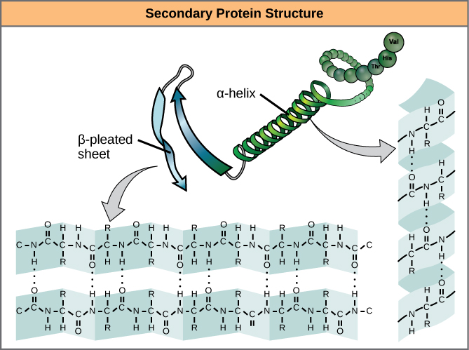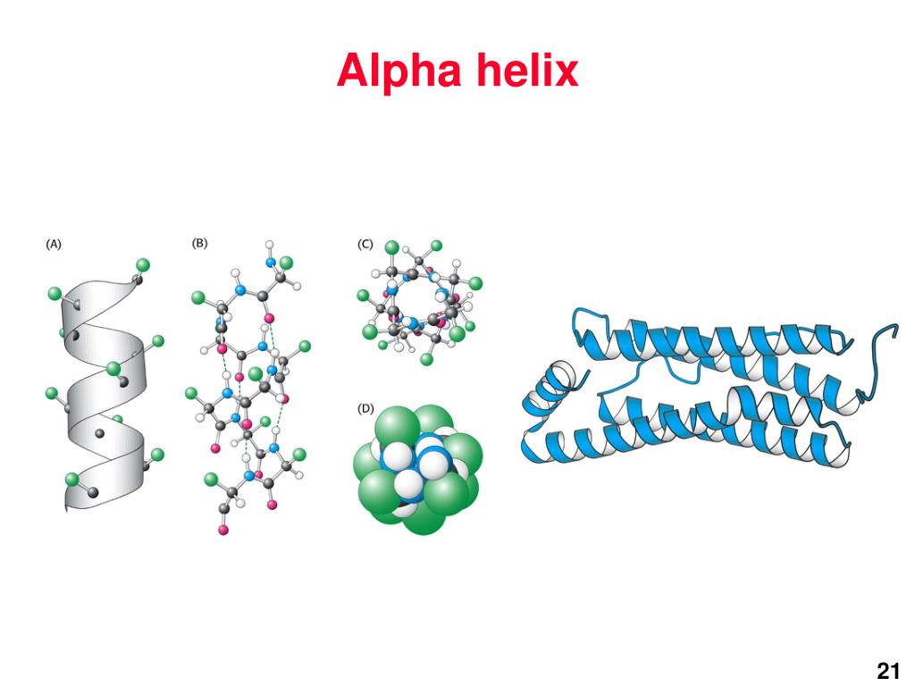
In the alpha helix, the bonds form between every fourth amino acid and cause a twist in the amino acid chain. Both structures are held in shape by hydrogen bonds. The most common are the alpha (α)-helix and beta (β)-pleated sheet structures. This can lead to a myriad of serious health problems, such as breathlessness, dizziness, headaches, and abdominal pain for those who have this disease.įolding patterns resulting from interactions between the non-R group portions of amino acids give rise to the secondary structure of the protein.

(credit: modification of work by Ed Uthman scale-bar data from Matt Russell)īecause of this change of one amino acid in the chain, the normally biconcave, or disc-shaped, red blood cells assume a crescent or “sickle” shape, which clogs arteries. In this blood smear, visualized at 535x magnification using bright field microscopy, sickle cells are crescent shaped, while normal cells are disc-shaped. The structural difference between a normal hemoglobin molecule and a sickle cell molecule-that dramatically decreases life expectancy-is a single amino acid of the 600.įigure 1. The molecule, therefore, has about 600 amino acids. What is most remarkable to consider is that a hemoglobin molecule is made up of two alpha chains and two beta chains that each consist of about 150 amino acids. In sickle cell anemia, the hemoglobin β chain has a single amino acid substitution, causing a change in both the structure and function of the protein. Any change in the gene sequence may lead to a different amino acid being added to the polypeptide chain, causing a change in protein structure and function. The unique sequence for every protein is ultimately determined by the gene that encodes the protein. The unique sequence and number of amino acids in a polypeptide chain is its primary structure. To understand how the protein gets its final shape or conformation, we need to understand the four levels of protein structure: primary, secondary, tertiary, and quaternary (Figure 2).

Folding can also result in covalent bonding between the "R" groups of cysteine amino acids.Due to protein folding, ionic bonding can occur between the positively and negatively charged "R" groups that come in close contact with one another.Hydrogen bonding in the polypeptide chain and between amino acid "R" groups helps to stabilize protein structure by holding the protein in the shape established by the hydrophobic interactions.

The amino acids with hydrophilic "R" groups will seek contact with their aqueous environment, while amino acids with hydrophobic "R" groups will seek to avoid water and position themselves towards the center of the protein. The "R" group of the amino acid is either hydrophobic or hydrophilic. Hydrophobic interactions greatly contribute to the folding and shaping of a protein.There are several types of bonds and forces that hold a protein in its tertiary structure. Tertiary Structure refers to the comprehensive 3-D structure of the polypeptide chain of a protein.


 0 kommentar(er)
